This example shows a mobile structure at the entrance of the left atrial appendage. With tissue velocity imaging, there is an alaising in the area of the highly mobile thrombus. After the injection of a contrast agent, the mobile thrombus is not visible. Therefore, a broad-based filling defect can be recognized, a “laminated” thrombus. Unfortunately, the contrast enhanced images have not been documented in the same planes as the unenhanced images. The term “laminated” thrombus is taken from the publication: Cardiovascular Imaging in the Management of Atrial Fibrillation, J Am Coll Cardiol 2006;48:2077– 84.

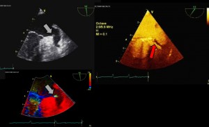
  

The non contrast enhanced images show a highly mobile thrombus at the entrance of the left atrial appendage.
Using the tissue velocity imaging, there is displayed an alaising, according to the high velocity of the mobile thrombus.
After the injection of microbubbles there is displayed a laminated thrombus, not seen on the non contrast enhanced images. Otherwise the highly mobile thrombus can not be further recognized, maybe due to the small size and the high mobility in relation to the frame rate.
Freezed frames of the mobile thrombus (grey arrows) and the laminated thrombus (red arrow), taken from the videos above (click to enlarge the picture).