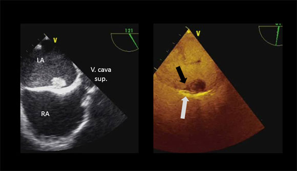
A patient with a CRT-D device has presented himself initialy with the clinical picture of a device infection 4 weeks ago. Device and electrodes have been removed and an antibiotic therapy has been initiated. The patient presents now with a worsening of heart failure without fever but slightly elevated leucocytes and CRP values. The video has been taken from a transesophageal echocardiography follow up study. Spontaneous echo contrast (SEC) in the left atrium has increased, matching a worsening of heart failure from NYHA II to NYHA III. Furthermore a spherical thrombus adheres next to the foramen ovale. Contrast administration (0.5 ml SonoVue) reveals a hypoechoic thrombus, which is not seen in the non-contrast-enhanced images as a result of spontaneous echo contrast (black arrow). Furthermore the “channel” of the foramen ovale is free of contrast signals as a sign of a thrombus (gray arrow).
| If the browser displayes the video distorted, please click on the white box next to the volume control of the player. The video is enlarged and by returning displayed in a non distorted version |
 |
|
