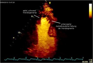
This video shows a severe apical perfusion defect during a stress echo with adenosine. A high degree stenosis of the left descending artery (LAD) was verified:
This video shows a short sequence, starting with a “flash”, followed by the replenishment of the microbubbles. There is a hyokinesia in the apex. The duration of the perfusion defect and the wall motion abnormality under stress indicates a hemodynamically significant high degree stenosis of the epicardial vessel. The endsystolic to early diastolic frame 4 seconds after flash reveals no perfusion signal in the apex (black area). There is a delayed replenishment in the anteroseptal wall segments. Ischemia is more pronounced in the apical und apical posterior segments, than in the septal segments.


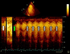
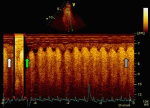
A so called “anatomical” m-mode is used to display perfusion during the flash sequence of the video above. In the apical anteroseptal wall segment there is a normal perfusion immedeatly before flash (white arrow). Immedeatly after flash the wall segment is completely black (green arrow), the bubbles are destroyed. The grey arrows point at a decreasing mainly subendocardial located perfusion defect. Replenishment is not completed within 5 seconds. The systolic wall thickening is reduced (hypokinesia) and a postsystolic shorting can be recognized.
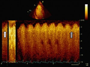
Blackening lasts longer in the apical posterior wall segment, caused by a much more pronounced ischemia. Systolic wall thickening is still normal.
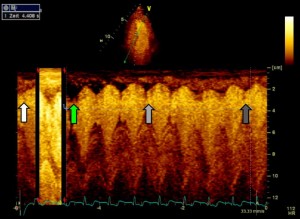
There is a transmural blackening during the first three cycles in the anterior apical wall segment (2-chamber view). Replenishment is afterwards not fully completed at the end of the recording. Wall “staining” is not the same at the point of the grey arrow compared to the white arrow. Easily to recognize the regular replenishment – within 1.5 seconds – of the mid inferior wall segment, which is strucked tangentially by the anatomical m-mode beam.
Even 6 seconds after flash at the end of the recording replenishment has not been completed (dark grey arrow) in the apical wall segments (3 chamber view). Please click an the images to enlarge.


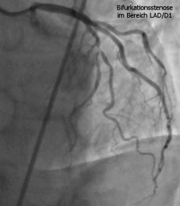
The angiogram reveals the expected high degree stenosis of the LAD and the first diagonal branch.